


?")
Pleurisy is the type of chest pain that occurs in pleura of the lungs when there is pleuritis. Since that may sound confusing—we will review each term. Read on to know what is pleuritis, and what experts like Pulmonologist in Lahore recommend for its management:
Table of Contents
What Are the Pleura?
Our lungs are surrounded by a thin layer of tissue, snug fitly in the chest cavity—known as the pleural membranes or the pleura. The pleural layers have space and fluid between them. The pleural membrane located close to the lungs is the visceral pleural membrane, while the outer layer is called the parietal pleural membrane.
Pleura diminish the friction between the lungs and the chest cavity during inhalation and exhalation. In normal conditions, these layers glide past each other—smoothly. However, in case of infection and inflammation, this movement is increasingly painful for the patient, particularly during deep breathing, sneezing, coughing and laughing
What is Pleuritis?
Inflammation and swelling of the pleura, secondary to any cause is called pleuritis. Pleuritis may be a complication of lung infection like pneumonia, or due to irritation of the pleura secondary to an autoimmune disorder—like rheumatoid arthritis, or lupus. Cases of chest trauma, as in contact sports or vehicular accidents can also result in pleuritis
Some cases of pleuritis are seen secondary to irritation of the pleura due to an embolus or a blood clot that travels to the lungs. Certain localized tumors, or metastatic diseases also causing irritation of the pleural membranes, and subsequent pain and inflammation.
How is Pleuritis Diagnosed?
Pleuritis is diagnosed with the help of imaging techniques like the following
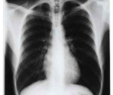
Chest x-Ray:
One of the first investigations ordered by the healthcare provider is the chest x-ray. Inflammatory changes in the lungs, as well as, fluid buildup in the chest are easily visible in this non-invasive investigation. It also shows space, gas or air between the lining, if present. The latter is called pneumothorax, which may be a complication of infectious lung disease.
CT Scan:
A CT scan uses x-ray radiation but builds a more detailed image of the lungs than a simple x-ray. A CT also helps to pinpoint the cause of pleurisy—especially in localizing the blood clot in the lung known as pulmonary embolism. Alternatively, when blood gets in the pleural space, it is easily picked up by a CT scan.
Blood Test:
Blood tests reveal infections in the body and is helpful in monitoring autoimmune disorders like lupus and rheumatoid arthritis.
Pleural Fluid Testing:
Fluid is taken from the subpleural space to test for the presence of inflammatory mediators, protein, or for presence of tumor cells. This process—also known as thoracentesis—also helps the patient breathe better.
Thoracoscopy:
To look inside the chest cavity, a thin, flexible tube known as a thoracoscope is used by the healthcare provider. This is particularly useful in case of diagnosing tumor cells.
How to Treat Pleuritis?
The treatment of pleuritis is centered around management of the underlying cause. If pleuritis is the consequence of any bacterial lung infection, the treatment option is antibiotics; similarly, for viral and fungal infections, antiviral and antifungal drugs are the treatment of choice. Along the antimicrobials, pain medication like NSAIDs—nonsteroidal anti-inflammatory drugs—and acetaminophen are used for discomfort.
Other treatment options include: prescription cough suppressants, to manage the cough, mucus breaking medication—known as mucolytics, medication to break blood clots if present, and bronchodilator inhalers to make breathing easier.
Thoracentesis is both a diagnostic tool and a treatment option. In case of large pleural effusions, the fluid is removed through thoracentesis, performed by or under the supervision of expert like Pulmonologist in Islamabad.